Practical Techniques for Classifying and Identifying Mica Ore Fragments
Mica ore fragments, derived from both primary mining and secondary processing, require precise identification for industrial applications. This guide outlines practical methods to distinguish between different types of mica fragments based on their physical, chemical, and structural properties.
Visual Inspection and Physical Properties
Color and Luster
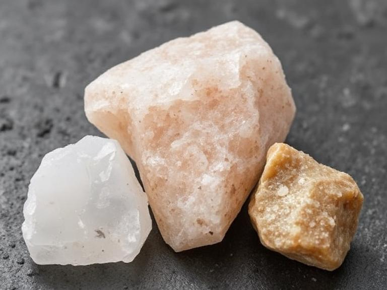
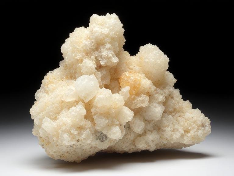
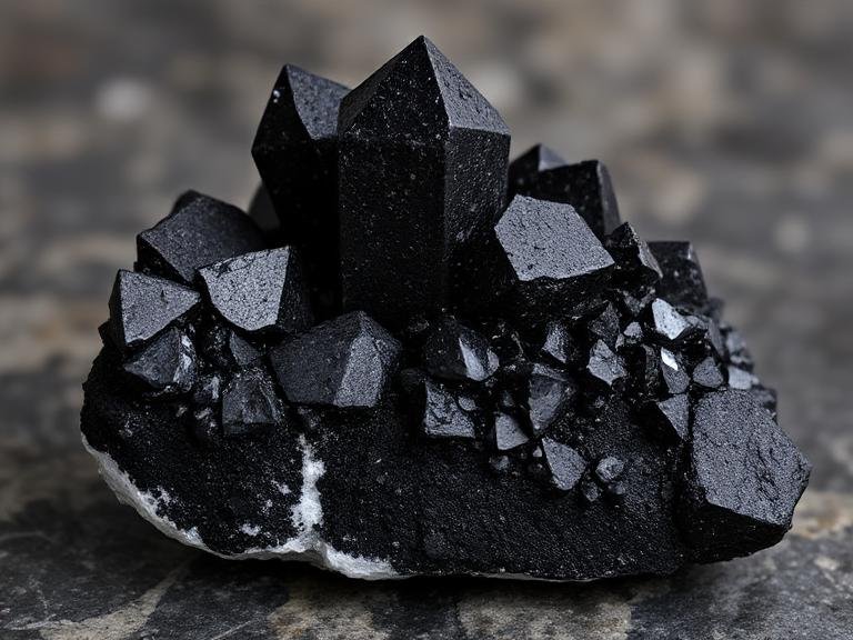
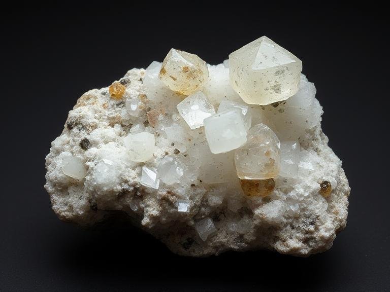
Mica fragments exhibit distinct colors depending on their mineral composition. Muscovite, a common variety, typically appears colorless to pale green or brown, with a vitreous to pearly luster. Biotite mica, on the other hand, is dark brown to black with a submetallic luster. Phlogopite, another variant, ranges from yellow to golden-brown, often displaying a similar luster to muscovite.
When inspecting fragments, note their overall color and how light reflects off their surfaces. Muscovite’s transparency or translucency, combined with its light color, helps differentiate it from biotite’s opaque, dark appearance. Phlogopite’s golden hue is a key identifier, especially when compared to the more subdued tones of muscovite.
Cleavage and Fracture
Mica minerals are renowned for their perfect basal cleavage, which allows them to split into thin, flexible sheets. During visual inspection, gently apply pressure to the edges of fragments using a knife or nail. Muscovite and phlogopite will split into smooth, flat sheets, while biotite may produce thinner, more brittle flakes due to its higher iron content.
Fracture patterns also provide clues. Mica fragments that break irregularly rather than along cleavage planes may indicate impurities or weathering. For instance, biotite fragments exposed to moisture often exhibit signs of alteration, such as a duller luster or a tendency to crumble rather than cleave.
Chemical and Structural Analysis
Acid Reaction Tests
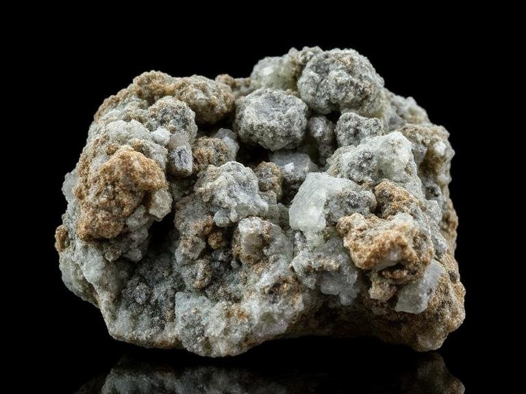
Certain mica varieties react predictably to acids, offering a simple yet effective identification method. Greenish muscovite mica carbonate schist, a subtype containing carbonate minerals, will fizz when exposed to dilute hydrochloric acid (HCl). This reaction occurs due to the release of carbon dioxide gas from carbonate components like calcite or dolomite.
To perform this test, place a small fragment in a dish and add a few drops of HCl. Observe for effervescence—a vigorous reaction suggests the presence of carbonate minerals, distinguishing this subtype from pure muscovite or biotite, which remain inert.
Microscopic Examination
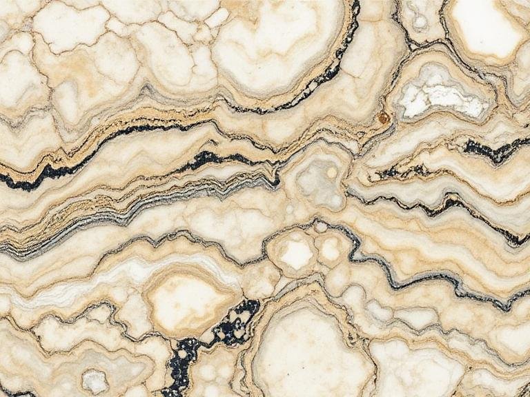
For more detailed analysis, a petrographic microscope reveals structural differences between mica types. Muscovite displays thin, colorless to pale-colored flakes with low birefringence under polarized light. Biotite, in contrast, appears darker and exhibits higher birefringence due to its iron and magnesium content. Phlogopite shares similarities with muscovite but often contains tiny inclusions or alterations that affect its optical properties.
When examining fragments, focus on their cleavage planes and inclusions. Muscovite’s clean, uniform cleavage contrasts with biotite’s often-disrupted planes caused by iron oxidation. Phlogopite may show signs of alteration, such as sericite (fine-grained muscovite) replacement, which appears as wispy, white streaks under magnification.
Advanced Identification Techniques
X-Ray Diffraction (XRD)
XRD analysis provides definitive identification by analyzing the crystalline structure of mica fragments. Each mica variety has a unique XRD pattern due to differences in atomic arrangement. For example, muscovite’s pattern features distinct peaks corresponding to its layered silicate structure, while biotite’s pattern includes additional peaks from iron and magnesium ions.
To use XRD, prepare a powdered sample of the fragment and analyze it using an X-ray diffractometer. Compare the resulting pattern to reference databases for mica minerals. This method is particularly useful for distinguishing between closely related varieties like phlogopite and eastonite, which may appear similar under a microscope but have distinct XRD signatures.
Infrared Spectroscopy (IR)
IR spectroscopy detects molecular vibrations in mica fragments, offering insights into their chemical composition. Muscovite produces characteristic peaks in the IR spectrum related to its hydroxyl (OH) groups and silicon-oxygen (Si-O) bonds. Biotite and phlogopite exhibit additional peaks due to iron and magnesium ions, altering their vibrational modes.
For IR analysis, prepare a thin film or KBr pellet of the fragment and scan it using an IR spectrometer. Compare the spectrum to known mica references to identify the specific variety. This technique is valuable for detecting minor components or alterations, such as the presence of chlorite in weathered biotite fragments.
Practical Applications in Industry
Quality Control in Mining and Processing
Accurate identification of mica fragments ensures consistent product quality in mining and processing operations. By distinguishing between high-grade muscovite and lower-quality biotite or phlogopite, producers can optimize sorting and blending processes. For example, muscovite fragments with high transparency and low iron content are preferred for electrical insulation applications, while biotite may be suitable for fillers or pigments.
Environmental and Safety Considerations
Identifying mica fragments also aids in environmental management and worker safety. Biotite fragments, which may contain higher levels of iron and other metals, require different handling procedures to prevent contamination or health risks. Similarly, recognizing weathered or altered mica varieties helps assess their stability in storage or transportation, reducing the likelihood of dust generation or structural failure.